Techniques of Safe Injections
Techniques of Safe Injections
- Right Methods of Injection Delivery
- Steps for administering the intradermal injection
- Consequence of unsafe intramuscular injection
- IV Cannulation
- Key aspects in IV Cannulation are
- Steps of inserting Catheter
- Securing and dressing the cannula
- Flushing IV Cannula
- Care of the catheter after insertion
- Blood collection could be of two types
Right Methods of Injection Delivery
Seven Rights for Safe Injection Delivery:- Right medication
- Right dose
- Right patient and site
- Right time
- Right route of administration
- Right documentation
- Right disposal
Injection can be delivered through different methods: The injections are commonly classified by the target tissue (e.g. intradermal, subcutaneous, intramuscular, intravenous, intraosseous, intra-arterial and peritoneal).
Intradermal Injection: A shallow injection given between the layers of the skin, creating a "weal" on the skin.

Steps for administering the intradermal injection
- Assemble equipment and check physicians older.
- Explain procedure to patient
- Perform hand wash and use disposable gloves.
- If necessary, withdraw medication from the ampoule or vial.
- Select an area on the inner aspect of the forearm that is not heavily pigmented or covered with hair. The upper chest or upper back beneath the scapulae are also sites for intradermal injections.
- Cleanse the area with an alcohol swab by wiping with a firm circular motion and moving outward from the injection site. Allow skin to dry.
- Use your non-dominant hand to pull skin taut over the injection site.
- Remove needle cap with non-dominant hand by pulling it straight off.
- Place the needle almost flat against the patient’s skin, bevel side up.
- Insert the needle so that the point of the needle can be seen through the skin--only about 1/8 of an inch.
- Slowly inject agent while watching for a small wheal or buster to appear. If none appears, withdraw the needle slightly.
- Withdraw the needle at the same angle it was inserted.
- Do not massage the area after removing the needle.
- Do not recap the used needle. Discard the needle and syringe in the appropriate receptacle.
- Assist the patient into a position of comfort.
- Remove your gloves and dispose them properly. Perform hand hygiene.
- Chart the administration of medication, as well as the site of the administration. Some agencies recommend circling the injection site with ink.
- Observe the area for signs of reaction at ordered intervals.
An intramuscular (IM) injection
The following are safe areas to give an IM injection:- VastusLateralis Muscle (Thigh): Look at your thigh and divide it into 3 equal parts. The middle third is where the injection will go. The thigh is a good place to give yourself an injection because it is easy to see. It is also a good spot for children younger than 3 years old.
- Ventrogluteal Muscle (Hip): Have the person getting the injection lie on his or her side. To find the correct location, place the bed of your hand on the upper, outer part of the thigh where it meets the buttocks. Point your thumb at the groin and your fingers toward the person's head. Form a V with your fingers by separating your first finger from the other 3 fingers. You will feel the edge of a bone along the tips of your little and ring fingers. The place to give the injection is in the middle of the V. The hip is a good place for an injection for adults and children older than 7 months.
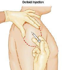
 Deltoid Muscle (Upper arm muscle): Completely expose the upper arm. You will give the injection in the center of an upside down triangle. Feel for the bone that goes across the top of the upper arm. This bone is called the acromion process. The bottom of it will form the base of the triangle. The point of the triangle is directly below the middle of the base at about the level of the armpit. The correct area to give an injection is in the center of the triangle, I to 2 inches below the acmmion process. This site should not be used if the person is very thin or the muscle is very small.
- Dorsogluteal Muscle (Buttocks): Expose one side of the buttocks. With an alcohol wipe draw a line from the top of the crack between the buttocks to the side of the body. Find the middle of that line and go up 3 Inches. From that point, draw another line down and across the first line, ending about halfway down the buttock, You should have drawn a cross. In the upper outer square you will feel a curved bone. The Injection will go in the upper outer square below the curved bone. Do not use this site for Infants or Children younger than 3 Years old. Their muscles are not developed enough. Even in adult patients, this site is best avoided as the extra layer of fat tissue reduces the absorption of the medication.
Consequence of unsafe intramuscular injection
An intramuscular injection could cause an infection, bleeding, numbness, swelling or pain.
Intra vascular with in blood vessel
Intravenous Injection is an injection given into a vein.
IV Cannulation
IV cannulation is an intravenous infusion (via a catheter placed in peripheral veins of upper limb)and is one of the commonest invasive procedures performed in acute care hospitals. The main indications of IV cannulation are:
- Fluid and/ or electrolyte replacement
- Route for drug administration
- Route for nutritional support
- Transfusion of blood and blood products
- Venous access for diagnostic blood draws
Key aspects in IV Cannulation are
- Selecting a right vein: When choosing an appropriate vein for venipuncture, many factors are considered including patient medical history, age, body size, weight, general condition and level of physical activity. the condition of patient's vein is also considered. Apart from the type of IV fluid or medications to be administered, expected duration of IV therapy should also be kept in mind. A cannula should not be placed in area of Localized oedema, Dermatitis, Cellulitis, Arteriovenous fistulae, Wounds, Skin grafts, Fractures, Stroke, Planned limb surgery and Site of previous cannulation.
- Selecting the right site: Site selection should be routinely initiated from distal to proximal.
- Selecting the right size of IV catheter
- 26 to 24 gauge for infants and children
- 24 22 gauge for children and elderly patients
- 24 20 gauge for medical patients and post “operative surgical patients
- 18 gauge for surgical patients and for rapid blood administration
- 16 gauge for trauma patients and those requiring large volumes of fluid rapidly.
- Preparing the site, inserting the catheter and right technique for its fixation: Select a vein, if the site is excessively hairy, the hair should be clipped, the site should not be shaved because it causes micro abrasions. Visibly dirty skin should be cleaned with soap and water. Then antimicrobial solution (Chlorhexidine gluconate) should be used. Tinctures of iodine 2%, 10% povidone-iodine, 70% isopropyl alcohol are also acceptable agents. Chlorhexidine gluconate achieves its antimicrobial action within 30 seconds while povidone iodine requires at least 2 minutes killing organisms on the skin. 70 % isopropyl alcohol should not be applied after a 10% povidone iodine preparation because this may irritate the skin and it interferes with povidone germicidal action. If a patient is allergic to iodine, the preparing solution of choice is chlorhexidine gluconate or 70% isopropyl alcohol.
Steps of inserting Catheter
Applying a tourniquet: The tourniquet is applied 2-3 inches above the intended venipuncture site, When tourniquet is in place, the patient should be asked to open and dose his fist several times to encourage venous distension. Gently rubbing or stroking the arm to warm the skin, and covering the entire arm with moist compresses also triggers the vasodilation. The vein should be palpated gently to see if it feels soft and bouncy.
Inserting the cannula: Before performing venipuncture, the vein should be stretched and immobilized Steps for inserting cannulae are
- The right hand should be used to grasp the cannula or the cannulas wings and proceed at once with venipuncture.
- The cannula should be inserted at 10 to 30 degrees angle, depending on the veins depth.
- The blood backflow in the cannula tubing or hub should be observed as it signifies that the cannula is in the vein lumen.
- Once the backflow has been observed, the cannula is lowered almost parallel to skin. The catheter is then pushed off the stellate and advanced completely into the lumen of the vein.
- Once the cannula is totally advanced into the vein the tourniquet is released and digital pressure is applied beyond the cannula tip and the hub stabilized.
Securing and dressing the cannula
- Tape placed under a transparent dressing should be clean. A ID 2- inch wide strip of tape is placed across the cannula hub: it should not cover the puncture site.
- Then ID 2 inch wide strip of tape is placed under the cannula hub, adhesive side facing up. The tape is then folded around the cannula hub. If the catheter hub with wings is being used, the tape strip is folded across the wings rather than the hub. The venipuncture site and catheter hub is covered with the dressing; the hub tubing junction is not covered. A gauze pad folded and covered with tape is placed under the cannula hub tubing junction to prevent the skin breakdown.
Flushing IV Cannula
- Regular flushing with 5 ml of sodium chloride 0.9% is sufficient, 2m1 before and 3m1 after administering the drug. Alternatively, safer solutions include using prefilled single use saline flush syringes.
Care of the catheter after insertion
- Documentation
- Site inspection
- Termination of infusion therapy
- Post removal
- Complications of IV Cannulation
- Phlebitis
Blood collection could be of two types
 Venous Blood collection-Venous blood collection is the method adopted for all needs of blood collection, where more than a few drops are required. The blood could be collected through traditional method of using a syringe and a needle or through a newer and much safe method of evacuated tubes. The clinical Laboratory recommendations approve dosed system of blood collection through evacuated tubes over the traditional method of drawing blood into a syringe and then shifting to the properly labeled testing container or tube. The collection sites should have adequate sharps management plan.
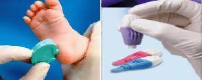
- Capillary blood collection- This is useful when only one to two drops of blood collection are required.

Last Modified : 2/21/2020
